 Skip to content
 Has this article been insightful? Share it!
Cerebrospinal fluid (CSF) is a clear, colorless fluid that surrounds the brain and spinal cord. It cushions the brain, regulates intracranial pressure, delivers essential nutrients, and removes metabolic waste.
Cerebrospinal fluid disorders occur when there is abnormal production, circulation, absorption, or leakage of CSF. These disorders may lead to increased intracranial pressure, brain fluid accumulation, or neurological symptoms such as headaches, vision problems, or cognitive decline. Early diagnosis and treatment are critical to prevent long-term complications.
A CSF leak occurs when cerebrospinal fluid escapes through a tear or defect in the dura mater, the protective membrane surrounding the brain and spinal cord.
CSF leaks may occur spontaneously due to skull base defects or high intracranial pressure, or they may follow trauma or surgery. Symptoms can also include nausea, dizziness, or sensitivity to light.
Cerebrospinal fluid disorders generally arise from two mechanisms: blockage of CSF pathways or impaired absorption of CSF into the bloodstream.
Patients experiencing persistent neurological symptoms may benefit from evaluation by a Singapore neurosurgeon for accurate diagnosis and treatment planning.
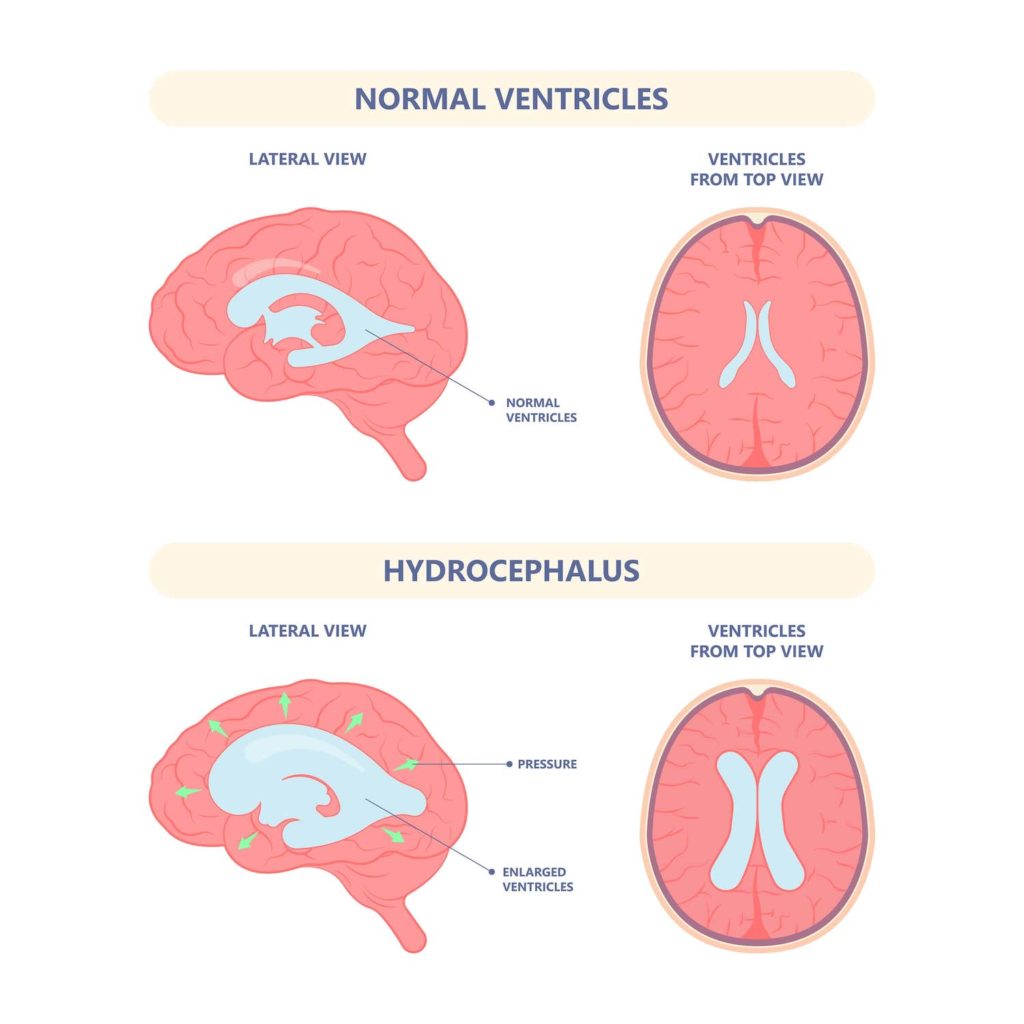
Hydrocephalus is the abnormal accumulation of cerebrospinal fluid within the brain’s ventricles, resulting in raised intracranial pressure.
This condition occurs when a structural blockage in the brain’s ventricles is present from birth, preventing normal CSF flow.
Communicating hydrocephalus occurs when CSF pathways remain open but absorption into the venous circulation is impaired.
Unlike obstructive hydrocephalus, there is no blockage within the ventricles. Instead, CSF accumulates due to absorption disruption.
Normal pressure hydrocephalus typically develops in individuals over 70 years old.
Despite enlarged ventricles, CSF pressure may remain within normal limits. Early recognition can improve management outcomes.
Seek medical attention if you experience:
Early assessment helps prevent life-threatening complications such as infection or acute intracranial pressure rise.
CSF disorders may result from:
Spontaneous CSF leaks may be linked to chronic intracranial pressure or underlying connective tissue abnormalities.
During the first consultation, your doctor would ask various questions to get a better understanding of your condition, such as:
These inquiries will be followed by a physical examination and various diagnostic tests to ensure an accurate diagnosis.
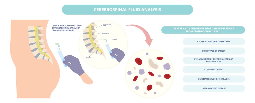
Diagnosis may include:
The prognosis depends on the underlying cause:
Treatment options include:
Shunt complications may include blockage or infection, which require timely follow-up.
Cerebrospinal fluid disorders are mainly related to CSF metabolism. The conditions are caused by any anomaly in CSF metabolism, which leads to excessive production or reduced absorption. As they can be life-threatening at times, these conditions should be properly diagnosed and treated as soon as possible to prevent harmful outcomes. Whether you are seeking consultation regarding CSF disorder symptoms or brain tumour treatments, do not hesitate to reach out to us today!
Cerebrospinal fluid disorders can significantly affect brain and spinal cord function if untreated. Accurate diagnosis and early intervention reduce the risk of infection, neurological decline, and permanent damage. Patients with symptoms suggestive of CSF leak or hydrocephalus should seek prompt evaluation.
If you are experiencing symptoms of a possible CSF leak or hydrocephalus, early evaluation is essential to prevent complications such as meningitis or permanent neurological damage.
Schedule a consultation
Senior Consultant
Neuro & Spine Surgeon
MBBS, MSc Surgery, MRCS (Edin),
MMed. Sc (Gen Surg) (S'pore),
FRCS. Surgical Neurology (UK)
Dr Sein Lwin is an experienced Senior Consultant Neuro & Spine Surgeon and the Neurosurgical Director at the Advanced Brain and Spine Surgical Centre.
He is highly experienced in spine surgery and in minimally invasive approaches for spinal cord tumours and degenerative spine surgery. His specialised interests lie in endoscopic endonasal and open skull base surgery, pituitary tumours, vascular surgery, cranial nerve disorders and peripheral nerve conditions.
Many neurological conditions may require urgent attention. If you require immediate care, please contact us.
Contact Us For More Information
We provide quality specialised care for neuro and spine conditions.
For enquiries, leave a message and our friendly team will get in touch with you.
Monday – Friday: 9:00AM – 5:00PM
Saturday: 9:00AM – 12:30PM
Sunday & Public Holiday: Closed
We provide quality specialised care for neuro and spine conditions.
For enquiries, leave a message and our friendly team will get in touch with you.
Monday – Friday: 9:00AM – 5:00PM
Saturday: 9:00AM – 12:30PM
Sunday & Public Holiday: Closed
We provide quality specialised care for neuro and spine conditions.
For enquiries, leave a message and our friendly team will get in
touch with you.
Monday – Friday: 9AM – 1PM | 2PM – 5PM
Weekends & Public Holidays: CLOSED
Member of Beyond Medical Group
© 2023 All Rights Reserved | Advanced Brain & Spine Surgical Centre | Terms & Conditions
 Optimized by Seraphinite Accelerator