Brain function requires constant oxygen and blood supply. Therefore, any disruption in these will severely affect brain function and cause severe complications — for example, stroke.
As blood vessels are responsible for the transportation of oxygen and blood supply, a blood vessel disorder is a serious condition that could potentially damage the rest of the body and be life-threatening.
Specifically, neurovascular conditions refer to all blood vessel disorders in the brain and spine where an area of the brain is temporarily or permanently affected by either bleeding or restricted blood flow.
Blood flow may be affected by:
● The narrowing of the vessel walls by hardening or abnormalities of the blood vessels
● A blockage caused by a clot or atheromatous plaque
● A ruptured blood vessel (haemorrhage)
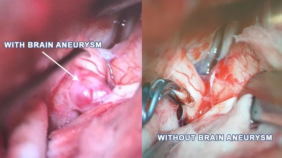
The ballooning of a blood vessel in the brain. The ballooning is caused by a weakened portion of the blood vessel wall, and this thinning may cause a rupture and bleeding.
A congenital abnormality in the formation between the arteries and veins which may lead to a bleed and rupture.
Also known as cavernous angiomas or cavernous malformations, these involve the enlargement and deformities of blood vessels clustering into an abnormal collection of thin-walled blood vessels.
Abnormal connections between an artery and vein within the venous sinus wall, which is the tough covering over the brain or spinal cord.
A blood clot that forms in the brain’s venous sinuses. This may cause a haemorrhage.
These conditions may cause a haemorrhage, bleeding or blockage, and may collectively cause a stroke.
Bleeding in or around the brain due to ruptured blood vessels can cause a stroke — this is termed a haemorrhagic stroke. There are two main types of haemorrhagic stroke:
● Bleeding within the brain
When an artery in the brain bursts and leaks out into brain tissue at high pressure, brain cells are killed or start to swell, and this is known as intracerebral haemorrhage.
● Bleeding on the surface of the brain
If blood vessels near the surface of the brain burst and leak into the subarachnoid space (a cushion of membranes that surrounds the brain), this is termed a subarachnoid haemorrhage.
Although there is no clear cause of neurovascular disease, certain conditions may put one at greater risk of developing it. These are:
● High Blood Pressure (Hypertension)
High pressure of blood flow can damage artery and blood vessel walls over time, increasing the chances of blood vessels bursting.
● Congenital blood vessel abnormalities
Rarely, people are born with abnormalities in their blood vessels. These vascular malformations are tangles of blood vessels or enlarged blood vessels which causes bleeding in the brain if vessel walls break.
● Atherosclerosis
The narrowing or “hardening” of the arteries happens when a fatty substance called plaque builds up inside the arteries. This is termed atherosclerosis and causes blood vessels to narrow, slowing or blocking the flow of blood. Smaller arteries in the brain are more prone to bleeding and could lead to haemorrhagic stroke.
● Cerebral Amyloid Angiopathy (CAA)
In CAA, a type of small vessel disease more common among older people with dementia, a protein called amyloid builds up inside the small blood vessels near the surface of the brain. The resulting damage can cause a vessel to tear, causing bleeding.
Risk factors may include diabetes, high blood pressure, high cholesterol, smoking, lack of physical activity, and genetic factors. Blood-thinning medication could also increase the risk of bleeding in the brain.
For haemorrhagic strokes, the FAST model is helpful in its detection.
● F – Facial weakness: Take note of drooping mouth or eyes
● A – Arm weakness: Check the mobility of both arms
● S- Speech problems: Is the speech slurred or incomprehensible?
● T- Time to call 999: if you see any of these signs.
● Arteriovenous malformation (AVM)
An AVM is a congenital defect between the arteries and veins. This condition affects the connection between these blood vessels and disrupts the flow of blood between them.
If the patient’s bleeding is severe, they may experience unbearable thunderclap headaches followed by a change of consciousness and coma.
Other general symptoms of blood vessel disorders could include:
● Sudden, intense headache
● Nausea and vomiting
● Stiff neck
● Blurred or double vision
● Sensitivity to light
● Seizures
● A drooping eyelid
● Loss of consciousness
● Problems with balance
It is advised to see a doctor at once if you have any signs or symptoms of blood vessel disorders, as they can be life-threatening and require emergency medical attention.
During your consultation, the specialist could assess your medical history and your symptoms, and conduct a physical examination that focuses on your brain and nerves. Medical tests ordered could include blood and urine tests and imaging tests.
Magnetic Resonance Imaging (MRI) is a test that uses powerful magnets, radio waves, and a computer to make detailed pictures, including that of soft tissues and the nervous system. The MRI protocol for stroke assessment is a group of MRI sequences put together to visualise the brain and blood vessel damage.
CT (computer tomography) cerebral venography is a contrast-enhanced examination with an acquisition delay, providing an accurate detailed depiction of the cerebral venous system, while the MR (magnetic resonance) venography uses magnetic resonance technology and intravenous (IV) contrast dye to visualize the veins.
Digital Subtraction Angiography (DSA) provides an image of the blood vessels in the brain to detect a problem with blood flow. The procedure involves inserting a catheter into an artery in the leg and passing it up to the blood vessels in the brain. A contrast dye is injected through the catheter and X-ray images are taken of the blood vessels.
Transcranial doppler (TCD) ultrasound is a test that uses sound waves to detect blood vessel disorders that affect blood flow in your brain. For example, stroke caused by blood clots, narrowed sections of blood vessels and tiny blood clots can be visualised.
CT perfusion and MRI perfusion imaging show which areas of the brain are adequately supplied or perfused with blood and provide detailed information on the delivery of blood or blood flow to the brain. Thus, these can be used to evaluate acute stroke and blood vessel constriction.
Treatment depends on the type of neurovascular condition you have. This could come in the form of medication and/or minimally invasive procedures such as:
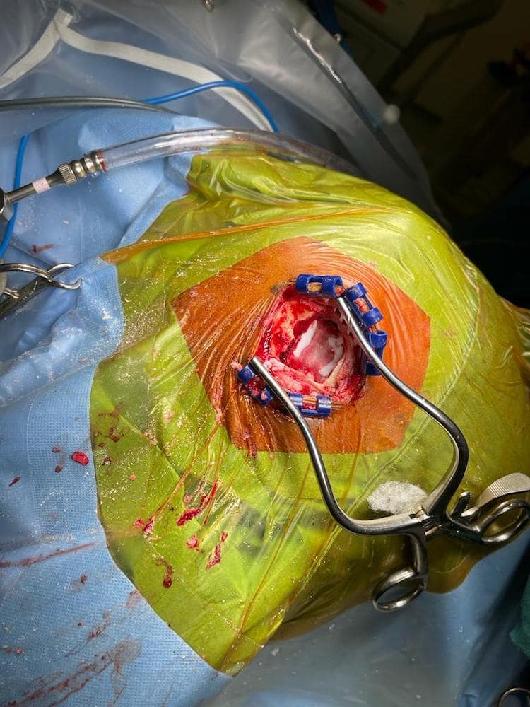
A bone flap of diameter 3-4cm in the skull is created in mini-craniotomy, and surgery is conducted to correct the aneurysm by placing coils of thin metal wires into the aneurysm. Blood clots that form around this coil prevent the aneurysm from rupturing.
A craniotomy is performed, where a part of the bone from the skull is surgically removed to expose the brain. The microscope is used to isolate and remove the AVM and cavernoma from the tissues around the brain.
Extracranial to intracranial (EC-IC) bypass surgery is performed to increase blood supply to the brain by surgically connecting the external carotid artery in your neck with the internal carotid artery inside your skull, thus bypassing the blocked area.
The inner lining of the carotid artery is surgically removed together with any obstructive deposits during endarterectomy, preventing narrowing (stenosis) of the blood vessel.
Our arteries play an integral part in transporting and supplying our brain and spine with nutrients and oxygen-rich blood. A neurovascular disorder leads to obstruction of the vessels that could cause serious disability or even death because of the restricted blood flow to the brain.
Treatment for blood vessel disorders often involves surgical intervention. If you or your loved one experiences signs and symptoms of a neurovascular dysfunction, please consult a neurosurgeon immediately.

Senior Consultant
Neuro & Spine Surgeon
MBBS, MSc Surgery, MRCS (Edin),
MMed. Sc (Gen Surg) (S'pore),
FRCS. Surgical Neurology (UK)
Dr Sein Lwin is an experienced Senior Consultant Neuro & Spine Surgeon and the Neurosurgical Director at the Advanced Brain and Spine Surgical Centre.
He is highly experienced in spine surgery and in minimally invasive approaches for spinal cord tumours and degenerative spine surgery. His specialised interests lie in endoscopic endonasal and open skull base surgery, pituitary tumours, vascular surgery, cranial nerve disorders and peripheral nerve conditions.
Many neurological conditions may require urgent attention. If you require immediate care, please contact us.
Contact Us For More Information
Your needs are important to us.
For enquiries, leave a message and our friendly team will get in touch with you.
Monday – Friday: 9:00AM – 5:00PM
Saturday: 9:00AM – 12:30PM
Sunday & Public Holiday: Closed
We provide quality specialised care for neuro and spine conditions.
For enquiries, leave a message and our friendly team will get in touch with you.
Monday – Friday: 9:00AM – 5:00PM
Saturday: 9:00AM – 12:30PM
Sunday & Public Holiday: Closed
We provide quality specialised care for neuro and spine conditions.
For enquiries, leave a message and our friendly team will get in
touch with you.
Monday – Friday: 9AM – 1PM | 2PM – 5PM
Weekends & Public Holidays: CLOSED
© 2023 All Rights Reserved | Advanced Brain & Spine Surgical Centre | Terms & Conditions